RECTAL NEUROENDOCRINE TUMORS
A rectal neuroendocrine tumor (or “NET”) is a growth arising from hormone-producing cells in the wall of the rectum. It is not the same thing as a typical colon cancer, and it does not behave in the same way.
Rectal neuroendocrine tumors are usually found by accident. Most are discovered during a colonoscopy done for screening or for another reason, appearing as a small, smooth, slightly yellow bump under the lining. They rarely cause symptoms of their own. The number diagnosed has risen sharply over the past thirty years, almost entirely because more people are having colonoscopy, and because the procedure is being performed with higher-resolution cameras that detect more subtle findings.

How rectal NETs behave
Most rectal neuroendocrine tumors are small and slow-growing, and the great majority never cause harm. About 90% are under 1 centimeter when found. At that size, and when the tumor is low grade and confined to the superficial layers of the wall, the chance that it has spread anywhere is very low, and long-term survival is the same as for people who never had one.
The word "tumor" can be confusing here. Small rectal NETs are officially classified as cancers, but biologically, a small, low-grade NET behaves nothing like a typical cancer. Small rectal NETs typically grow very slowly, and most never invade or spread. The "cancer" label reflects that these cells have the potential to behave aggressively. A small, low-grade NET that's been completely removed has a superb prognosis, and for most patients, no further treatment is needed.
That said, behavior is not uniform, and three things determine it: size, grade (how quickly the cells are dividing, measured on the removed tissue), and depth (how far into the wall it has grown). These findings are determined by our pathology samples when the lesion is biopsied or removed.
Why we remove rectal NETs
Even though most rectal NETs behave harmlessly, the standard approach for small ones is removal rather than watching. The reasoning is straightforward: removal is a short, fairly low-risk endoscopic procedure, it gives a definitive answer about grade and depth that no biopsy alone can provide, and it takes the question off the table permanently. Watching offers no comparable advantage.
Additionally, removing rectal NETs while small is more straightforward, while a larger one may require an operation.
Size, and what it changes
Under 1 centimeter. The most common situation, and the most reassuring one. Endoscopic removal is the standard treatment. If the tumor is 5 mm or larger, an endoscopic ultrasound from inside the rectum is often done first to confirm it has not grown deeper than expected.
Between 1 and 2 centimeters. An intermediate zone. Imaging is done first, typically endoscopic ultrasound from inside the rectum, plus or minus additional scans of the abdomen. If everything is confined and low grade, these can still often be removed endoscopically or through a minimally invasive surgery. Follow-up afterward is closer.
Over 2 centimeters. Less common, and more concerning. The risk of spread to nearby lymph nodes is substantial. These rectal NETs are discussed in a multidisciplinary meeting, and surgery to remove that part of the rectum, along with the lymph nodes, is usually recommended.
Grade and depth can override size. A small tumor that is high grade, or that has grown into the muscle layer, or that shows invasion of small blood or lymph vessels, is managed as a higher-risk lesion regardless of how small it is, and is discussed with a surgeon.
How removal is done
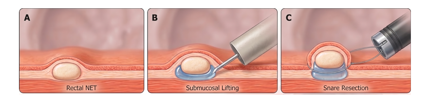
Most rectal NETs sit in the layer just beneath the surface lining of the rectum, which is why an ordinary snare polypectomy often leaves tumor behind at the deep edge. Techniques that capture that deeper layer are used instead. All are done through the colonoscope, under sedation.
Modified endoscopic mucosal resection. This is the most common approach for small rectal NETs. Fluid may be injected underneath to lift the lesion, and a cap or a rubber band device on the end of the scope may be used to draw the tumor and the tissue beneath it up before it is cut away with a wire snare. This is quick and highly effective.
Endoscopic submucosal dissection (ESD). A fine electrical knife is used to dissect the tumor off the wall in one intact piece. This takes considerably longer, and is favored for larger lesions where the margins matter more.
Full-thickness resection. A device that removes the whole thickness of the wall, sealing it with a clip. Used in selected cases, particularly where scarring from a previous attempt prevents the lesion from lifting.
If it has already been partly removed
Some of these are found only because a small bump was snared as if it were an ordinary polyp, with the neuroendocrine diagnosis coming back from pathology afterward. This is common and not a mistake, but it often means the deep margin is unclear. In that situation, a repeat colonoscopy and/or ultrasound is usually done to inspect the site and to take deeper samples, to ensure any remaining tumor is removed properly.
Risks of the rectal NET removal
Small, and comparable to other endoscopic resections. Bleeding afterward is the most common problem and is usually managed endoscopically. Making a hole in the wall is very uncommon and is generally closed with clips at the time. Rarely, surgery can be required to fix a hole if the area is too large to fix during endoscopically.
Follow-up
This depends entirely on the pathology results.
Small, low-grade, completely removed. Follow-up is individualized, but for a tumor under 1 cm removed with clear margins, several international guidelines advise no specific ongoing surveillance for the tumor itself, and no routine scans. Colonoscopy then follows the standard schedule that would apply to anyone. Recurrence in this group is rare. We often consider a single one year follow-up procedure at BIDMC as a last check of the area.
Between 1 and 2 centimeters, completely removed. Closer follow-up, with annual endoscopy for around five years commonly recommended, since the risk of later recurrence in this group is meaningfully higher.
Higher grade, deeper invasion, or larger tumors. Longer-term follow-up with scans as well as endoscopy, planned individually.
Suggested reading
The Neuroendocrine Tumor Research Foundation maintains plain-language patient information on gastrointestinal neuroendocrine tumors, including rectal tumors and what test results mean: netrf.org/for-patients